Aller au contenu

Modern medical imaging plays a vital role in patient care. It helps doctors see inside the body without surgery. This is especially true for conditions like high blood pressure and cardiovascular illness. These conditions affect millions of people worldwide.
Advanced scanning techniques have changed how we find health problems. They allow for very detailed pictures of the heart and the arteries leading to the kidneys. This helps spot issues early, often before a person feels sick. Early detection is key to better health outcomes.
This guide will explore these important imaging methods. We will cover how they help in the diagnosis and management of these interconnected conditions. Our focus is on a complete view of the patient’s health.
At Centre de Radiologie Diouri Kénitra, we use the latest technology. Our team is dedicated to providing precise assessments. We support clinicians across Morocco with clear, actionable results.
Key Takeaways
- Advanced imaging is crucial for detecting and managing high blood pressure and cardiovascular conditions.
- Modern techniques provide detailed visuals of the heart and kidney arteries for early problem identification.
- Early detection through scans can lead to better management before symptoms become severe.
- A comprehensive evaluation often requires looking at both cardiac and renal vascular health.
- Specialized radiological assessment is essential for identifying secondary causes of high blood pressure.
- State-of-the-art imaging services support informed clinical decisions for patient care.
Introduction to Radiology for Hypertension and Heart Disease
Medical scanning technologies have revolutionized how we approach blood pressure-related health concerns. Elevated blood pressure stands as a major preventable cardiovascular risk factor worldwide.
In the United States, nearly half of adults face this condition. European prevalence reaches about 25%. Control rates remain concerning, with one-quarter of affected individuals experiencing uncontrolled levels.
Uncontrolled elevated pressure damages vital organs. The heart, kidneys, brain, and vascular system suffer most. Early detection through imaging prevents serious complications.
We utilize various scanning methods for evaluation. Basic chest X-rays provide initial assessment. Advanced cross-sectional techniques offer detailed anatomical views.
| Region | Prevalence Rate | Control Status | Population Affected |
|---|---|---|---|
| United States | 47% | 75% Controlled | Adult Population |
| Europe | 25% | 75% Controlled | Adult Population |
| Global Average | 30-35% | Variable | Adults Worldwide |
Timely identification of organ damage significantly improves outcomes. It allows intervention before irreversible changes occur. Our approach focuses on comprehensive assessment for better management.
Accurate visualization guides therapeutic decisions effectively. It helps physicians identify secondary causes and monitor treatment progress through sequential studies.
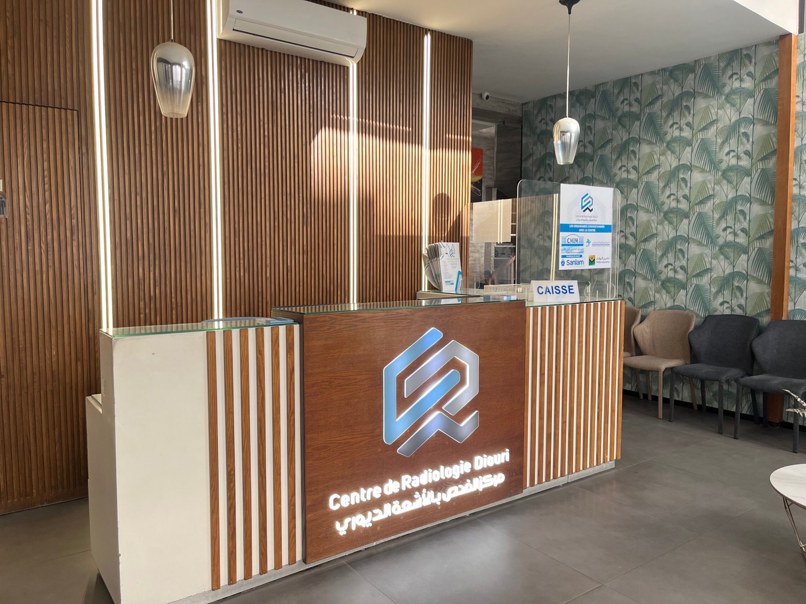
Overview of Centre de Radiologie Diouri Kénitra in Morocco
Centre de Radiologie Diouri Kénitra stands as a premier medical facility in northwestern Morocco. Our center provides advanced diagnostic services to the local community. We focus on delivering precise imaging results for better health outcomes.
Our team includes internationally trained specialists with expertise in cardiovascular and renal assessment. We utilize state-of-the-art equipment for accurate diagnosis. Each examination is conducted with the highest standards of safety and comfort.
We offer comprehensive services including computed tomography and echocardiography. Our specialized protocols are designed for detailed evaluation of vascular and cardiac health. This allows for thorough assessment of complex medical conditions.
Our approach prioritizes patient comfort and clear communication. We work closely with referring physicians across Morocco. Our detailed reports integrate clinical data with imaging findings for optimal patient management.
| Service | Purpose | Specialization |
|---|---|---|
| Computed Tomography | Vascular Assessment | Artery Imaging |
| Echocardiography | Cardiac Evaluation | Heart Function |
| Specialized Protocols | Detailed Examination | Organ Damage Detection |
We continuously invest in staff training and technology upgrades. This ensures our center remains at the forefront of diagnostic services in Morocco. Our commitment is to provide accessible, high-quality care for all patients.
Understanding Hypertension and Cardiac Disease in Radiologic Terms
The heart undergoes distinct structural changes when faced with persistent arterial pressure challenges. We define hypertensive heart disease as a spectrum of cardiac abnormalities developing from chronic pressure elevation.
Left ventricular hypertrophy represents the most common manifestation. This thickening of the heart muscle serves as an adaptive response to minimize wall stress. However, it eventually leads to impaired function if pressure remains uncontrolled.
When blood values exceed normal ranges, the risk of heart failure increases dramatically. Research shows approximately two-fold higher risk in male patients and three-fold in. Early radiological detection becomes critically important for prevention.
Different pressure patterns produce varying cardiac damage levels. All require careful assessment to determine true organ involvement extent.
| Hypertension Pattern | Cardiac Impact | Detection Priority |
|---|---|---|
| Sustained | Progressive LVH | High |
| White-Coat | Variable Changes | Moderate |
| Nocturnal | Diastolic Dysfunction | High |
Understanding radiologic terminology and imaging patterns associated with arterial hypertension is essential. These findings guide treatment intensity and help predict cardiovascular risk in individual patients.
The Role of Advanced Imaging in Diagnosing Cardiovascular Conditions
Contemporary medical visualization methods offer unprecedented insights into the workings of the cardiovascular system. These technologies have transformed how we detect and manage circulatory conditions.
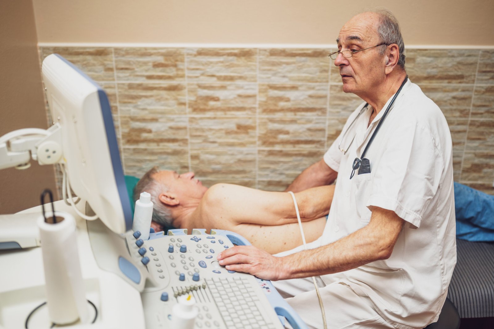
Echocardiography has long served as the primary tool for cardiovascular assessment. It provides real-time views of cardiac structures and function. This non-invasive approach allows early disease detection before symptoms appear.
Recently, cardiovascular magnetic resonance imaging has gained prominence. It offers unrestricted field views and detailed tissue characterization. This advanced imaging technique enables comprehensive evaluation of anatomical changes.
We select modalities based on clinical questions and patient needs. Each diagnosis requires careful consideration of available resources. Our goal is maximum information with minimal inconvenience.
Multiple imaging approaches often provide the most complete picture. They assess both structural and functional parameters simultaneously. This integrated assessment helps characterize the full extent of cardiovascular disease.
These technologies also track treatment effectiveness over time. They provide objective metrics that guide therapy adjustments for patients. This ongoing monitoring benefits patients throughout their care journey.
Imaging Techniques: Computed Tomography and Echocardiography
Computed tomography and echocardiography represent cornerstone modalities in modern cardiovascular diagnostics. These complementary approaches provide detailed anatomical and functional information.
Computed Tomography for Vascular Assessment
Computed tomography offers exceptional spatial resolution for vascular assessment. This imaging technique detects specific findings indicating pressure overload.
Key measurements include main pulmonary artery enlargement and right ventricular changes. The axial view reveals critical ratios like RV/LV >1. These findings help identify pulmonary vascular conditions.
Our computed tomography assessment extends throughout the vascular system. We evaluate renal arteries, coronary vessels, and pulmonary circulation comprehensively.
Echocardiography in Detecting Heart Abnormalities
Echocardiography provides real-time evaluation of cardiac function. This ultrasound-based imaging modality assesses chamber dimensions and wall motion.
Two-dimensional echocardiography calculates left ventricular volume using established formulas. Three-dimensional techniques eliminate geometric assumptions for greater accuracy.
We utilize echocardiography to monitor heart function over time. The short-axis view provides essential data on ventricular volume and ejection fraction.
Both computed tomography and echocardiography offer distinct advantages. We select the optimal imaging approach based on each patient’s clinical needs.
Step-by-Step How-To Guide for Heart Imaging Procedures
Our clinical practice emphasizes a structured methodology for cardiovascular assessment procedures. This systematic approach ensures consistent, high-quality results for every patient. We follow established protocols that have proven effective in clinical settings.
Patient preparation forms the foundation of successful imaging. We gather comprehensive medical history and review previous studies. Current medications and specific clinical questions guide our examination strategy.
The actual imaging process follows a careful sequence. For echocardiography, proper patient positioning in left lateral decubitus position is crucial. We systematically acquire standard views including parasternal long-axis and apical four-chamber images.
Computed tomography requires different preparation steps. Patients typically fast before the procedure and receive intravenous access. Contrast timing and breath-hold instructions are optimized for each individual.
Interpretation follows a methodical pattern. Radiologists initially survey for gross abnormalities before detailed analysis. They evaluate each cardiac chamber systematically, measuring dimensions and assessing wall motion.
We never disregard incidental findings during examination. Subtle abnormalities may indicate significant conditions requiring further assessment. Our comprehensive cardiac ultrasound guide provides additional technical details.
Time management balances thoroughness with efficiency. Most echocardiographic examinations require 30-45 minutes, while CT studies typically take 10-15 minutes. Interpretation time varies based on case complexity.
Quality assurance completes the process. We verify that all required images are obtained and measurements fall within expected ranges. The final report directly addresses the referring physician’s clinical questions.
Evaluating Kidney Arteries in Hypertensive Patients
Kidney artery evaluation provides crucial insights into secondary hypertension cases. Narrowed renal arteries can trigger persistent blood pressure issues. This condition demands specialized imaging approaches.
Renovascular hypertension accounts for significant secondary cases. Arterial narrowing reduces blood flow to kidneys. This activates the renin-angiotensin system, elevating pressure.
Our diagnostic approach begins with screening tests. Renal ultrasound with Doppler detects flow abnormalities. More definitive imaging follows with CT or MR angiography.
Classic findings include kidney size differences. The affected kidney often appears smaller. Delayed contrast excretion and visible stenosis confirm the diagnosis.
| Pathology Type | Affected Area | Patient Profile |
|---|---|---|
| Atherosclerotic Disease | Proximal Renal Artery | Older patients with risk factors |
| Fibromuscular Dysplasia | Mid-to-Distal Artery | Younger patients, especially women |
We employ urography with urea wash-out for functional assessment. Captopril renography shows asymmetric renal function. Direct arteriography provides detailed arterial visualization.
Identifying renovascular causes is vital. Many cases are potentially curable. Interventions like angioplasty or stenting can restore normal blood flow.
At our center, we use systematic protocols. We focus on patients with resistant arterial hypertension. Sudden worsening of control warrants thorough renal artery assessment.
Assessing Left Ventricular Hypertrophy and Cardiac Remodeling
Cardiac adaptation to stress involves distinct structural changes within the left ventricle. These changes are not just anatomical but carry significant implications for patient health.

We define left ventricular hypertrophy as an increase in left ventricular mass with parallel thickening of the wall. This represents the heart’s adaptive response to chronic pressure or volume overload.
Identifying Concentric and Eccentric Patterns
Our imaging analysis classifies these adaptations into specific patterns. Concentric hypertrophy occurs when left ventricular mass enlarges and walls thicken in response to pressure overload. This pattern often relates to lower cardiac output.
In contrast, eccentric hypertrophy shows increased left ventricular mass with chamber dilatation. It maintains normal relative wall thickness. This pattern is more common in young adults and correlates with higher cardiac output.
A third pattern, concentric remodeling, features normal left ventricular mass but increased relative wall thickness.
We utilize precise imaging parameters to distinguish these patterns:
- Measurements of interventricular septal thickness
- Posterior wall thickness
- Left ventricular internal dimensions
- Calculation of relative wall thickness
Identifying the specific pattern is clinically significant. Concentric hypertrophy carries higher risk for cardiovascular events compared to concentric remodeling. This knowledge guides treatment intensity for patients.
In China, asymptomatic left ventricular hypertrophy prevalence approaches 30% among patients with elevated blood pressure. Systematic screening helps detect this silent condition before dysfunction develops.
Accurate assessment requires high-quality imaging with precise measurements. At our center, we provide this expertise through standardized protocols. Early detection of these patterns enables timely intervention to preserve cardiac function.
Recognizing Radiologic Signs of Pressure Overload and Fibrosis
Advanced imaging reveals critical structural clues that signal underlying pressure overload and developing fibrosis within the heart muscle. Identifying these signs early is crucial for effective intervention.
We look for specific findings on computed tomography scans. Main pulmonary artery enlargement beyond 30 mm is a key marker. Right ventricular dilatation and a flattened septum also indicate elevated pressures.
Key Imaging Markers and Measurements
These signs manifest differently depending on the source of pressure. Left-sided overload often causes concentric wall thickening. Right-sided issues lead to chamber enlargement and septal bowing.
Myocardial fibrosis is a serious consequence. It results from excessive scar tissue deposition. This process causes progressive cardiac stiffness and dysfunction.
This fibrosis is an independent prognostic marker. Its presence signals higher risk for heart failure and other major events. Early detection through advanced techniques is vital.
Specific measurement thresholds help us assess the severity. We evaluate atrial dimensions for elevated atrial pressure. Increased wall thickness beyond normal ranges is another critical measurement.
Recognizing these findings allows for timely treatment intensification. This can potentially reverse remodeling and prevent symptomatic disease progression.
Integrating Patient History with Radiologic Findings
Effective medical assessment relies on weaving together a patient’s complete story with the visual evidence from scans. We never interpret imaging results in isolation. The clinical background provides essential context for accurate diagnosis.
Our systematic approach begins with the referring physician’s specific questions. We consider presenting symptoms like chest discomfort or breathing difficulties. Physical examination results guide our focused image interpretation.
Detailed patient history helps distinguish between conditions with similar appearances. Medication records are particularly important. Certain blood pressure drugs can reduce heart muscle thickness over time.
We review previous imaging studies when available. Comparison shows whether changes are stable or progressive. This temporal perspective is invaluable for treatment decisions.
At our center, we maintain open communication channels with referring physicians. This ensures our interpretations directly address each patient’s management needs. The integration of clinical and imaging data leads to optimal care.
Practical Steps in Conducting a Radiologic Examination
Precise execution of radiologic procedures requires careful coordination from preparation through documentation. Our systematic approach ensures each examination delivers optimal diagnostic value while maintaining patient comfort.
We begin with thorough patient preparation. This includes scheduling considerations and pre-procedure instructions. Our team verifies medical history and confirms examination indications.
Patient positioning is critical for quality imaging. For cardiovascular studies, we use specific techniques to optimize the diagnostic view. Proper positioning minimizes artifacts and enhances image clarity.
During the examination, we maintain real-time quality control. Our technologists immediately assess image quality and repeat suboptimal acquisitions. This proactive approach prevents the need for patient recalls.
We prioritize time-efficient workflows without compromising quality. Most cardiovascular imaging studies complete within 30-60 minutes. This balance respects our patients‘ schedules while ensuring comprehensive assessment.
Documentation completes the process. We record technical parameters and contrast volumes administered. All images are properly archived for future reference and comparison studies.
Interpreting Imaging Results for Optimal Diagnosis
Our systematic approach to analyzing diagnostic images ensures comprehensive assessment. We begin by verifying image quality and technical adequacy before proceeding with detailed evaluation.
Cardiac chamber sizes are measured against established reference ranges. These measurements account for body surface area, age, and gender. Normal left ventricular configuration appears circular to elliptical on short-axis view.

Septal flattening or leftward bowing indicates pressure overload. This finding suggests right ventricular dilatation. Such evidence points toward pulmonary conditions requiring attention.
We assess systolic function using ejection fraction measurements. Values below 50% indicate impaired contractility. Regional wall motion analysis helps distinguish between different cardiomyopathy types.
Diastolic function evaluation employs mitral inflow Doppler patterns. The E/A ratio provides crucial evidence of relaxation abnormalities. Reduced ratios signal grade I diastolic dysfunction.
| Function Type | Key Parameter | Abnormal Finding | Clinical Significance |
|---|---|---|---|
| Systolic Function | Ejection Fraction | <50% | Impaired contractility |
| Diastolic Function | E/A Ratio | Reduced | Relaxation abnormality |
| Ventricular Geometry | Septal Position | Flattening/Bowing | Pressure overload |
Our interpretation process includes peer consultation for complex cases. We compare current studies with prior examinations to track changes. This comprehensive evaluation supports accurate diagnosis and optimal patient management.
Comprehensive Radiology for Hypertension and Heart Disease Overview
The synergy between clinical findings and advanced visualization techniques enhances diagnostic precision. Our approach integrates multiple modalities into a unified framework.
Correlating Clinical Data with Imaging Insights
Echocardiography remains the cornerstone for assessing hypertensive cardiovascular conditions. It offers accurate evaluation of left ventricular geometry and function.
Cardiovascular magnetic resonance provides superior reliability for quantifying ventricular mass. This modality enables detection of subtle myocardial changes.
We systematically correlate blood pressure data with imaging observations. This integration helps determine disease severity and guide treatment decisions.
| Imaging Modality | Primary Strength | Clinical Application | Follow-up Utility |
|---|---|---|---|
| Echocardiography | Functional assessment | Initial evaluation | Portable monitoring |
| Cardiac MRI | Tissue characterization | Detailed analysis | Progression tracking |
| CT Angiography | Vascular anatomy | Comprehensive view | Surgical planning |
Serial imaging plays a crucial role in monitoring therapeutic response. Standardized protocols ensure consistent measurement of ventricular parameters over time.
At Centre de Radiologie Diouri Kénitra, we provide comprehensive services addressing all aspects of cardiovascular assessment. Our team collaborates closely with referring physicians to optimize patient management.
« The integration of clinical data with advanced imaging insights transforms how we approach complex cardiovascular conditions. »
This holistic approach enables early detection of complications like atrial enlargement. It supports personalized treatment strategies for better long-term outcomes.
Innovations and Future Trends in Cardiovascular Imaging
Emerging technologies are reshaping how we visualize and understand cardiac conditions. Cardiovascular magnetic resonance imaging offers revolutionary capabilities with unrestricted field views.
This modality provides superior tissue characterization for detailed assessment. It detects subtle disease processes that traditional methods might miss.
CMR excels at tracking left ventricular mass changes during follow-up periods. Its exceptional reproducibility enables detection of small variations as little as 5%.
Myocardial strain analysis can reveal asymptomatic ventricular dysfunction before ejection fraction declines. This allows earlier intervention for patients.
The extracellular volume fraction measured by CMR serves as an independent prognostic marker. It quantifies diffuse myocardial fibrosis for better risk stratification.
Artificial intelligence algorithms now automate image analysis and improve measurement consistency. These tools detect patterns not readily apparent to human observers.
Echocardiography continues evolving with 3D speckle tracking and contrast-enhanced studies. Handheld devices bring sophisticated cardiac imaging to point-of-care settings.
Future trends include hybrid imaging that combines anatomical and functional information. Molecular imaging visualizes disease processes at cellular levels.
We remain committed to incorporating validated advances into our evaluation protocols. This ensures patients receive the most sophisticated cardiovascular assessment available.
Strategies for Effective Patient Management and Follow-Up
We implement structured management strategies that prioritize ongoing assessment and timely intervention. Our approach begins with comprehensive baseline imaging studies. These initial scans document existing organ damage from elevated blood pressure.
Lowering systolic blood pressure significantly predicts left ventricular hypertrophy regression. Successful pressure control enables reversal of pathological cardiac remodeling. This makes aggressive management essential for better cardiovascular outcomes.
Reverting left ventricular mass through medication provides meaningful clinical benefits. Studies show reduced risk of stroke, myocardial infarction, and all-cause mortality. Serial imaging validates the value of LV mass reduction as a therapeutic target.
Improvement in global longitudinal strain relates primarily to LV mass reduction rather than blood pressure changes alone. Young adults often respond better to therapy than older patients. This demographic difference guides our treatment expectations and follow-up intervals.
We recommend repeat imaging at 6-12 month intervals for established cases. Extended intervals suit patients with mild disease and well-controlled pressure. Our multidisciplinary team ensures findings translate into appropriate therapeutic adjustments.
Conclusion
The integration of clinical data with imaging findings represents a powerful approach to cardiovascular care. This synergy enables early diagnosis before symptoms appear, reducing risk for patients.
Various imaging techniques provide objective evidence of organ damage. They track treatment effectiveness through serial evaluation. This monitoring has significant value in managing complex conditions.
Early detection of subtle signs allows timely intervention. This prevents progression to symptomatic disease. Comprehensive assessment guides personalized strategies.
At Centre de Radiologie Diouri Kénitra, we remain committed to excellence. Our advanced technology and experienced team support optimal care throughout Morocco. We invite collaboration for better cardiovascular health outcomes.